News
Anatomical Society Best Image Prize Awardees (May 2025)
Artwork Category:
Winner: Professor William A. Harris from the University of Cambridge.
Runner-Up: Dr Jennifer Z. Paxton from the University of Edinburgh.
Scientific Category:
Winner: Professor Abigail Tucker from King's College London.
Runner-Up: Mr Juan Carlos Palomeque from the Royal College of Surgeons in Ireland.
Please read the information below to find out more about the award-winning images. To view the images please click on the blue text next to the image title.
Best Image Prize (Artwork Category)
|
Image Title |
|
|
Description |
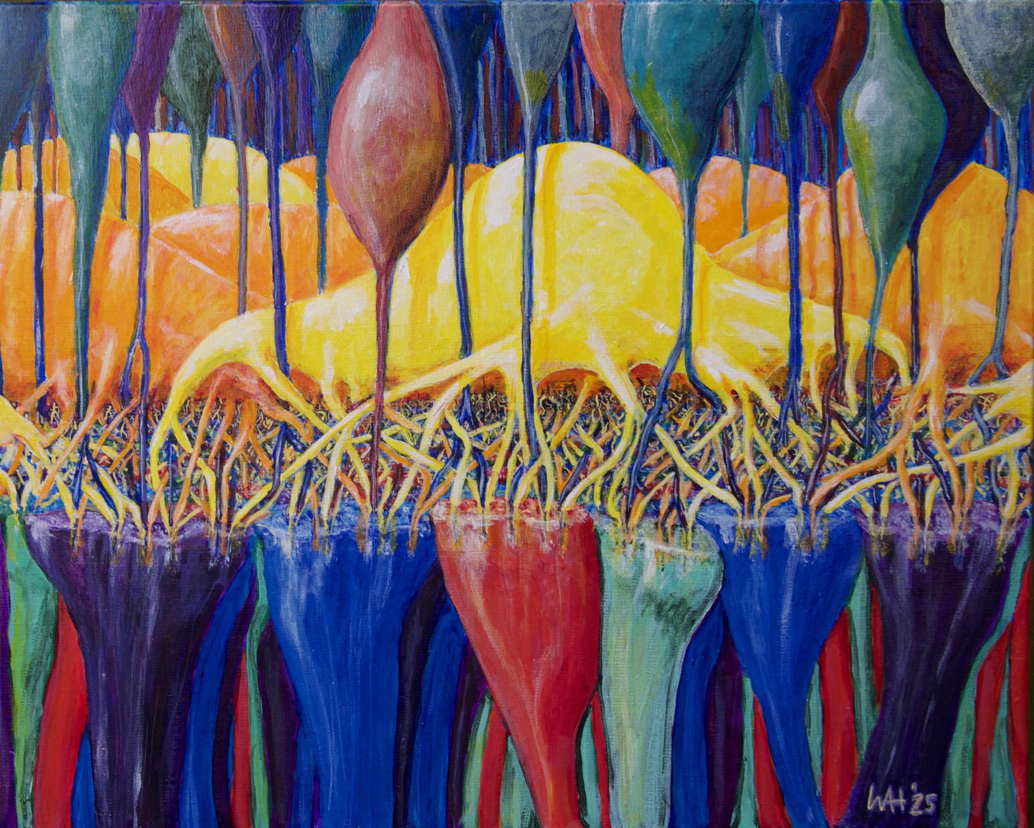
Pedicles of 4 type of cones (UV Blue Green Red, bottom), 3 types of Horizontal Cell (H1 yellow, H2 and H3 orange), Many types of Bipolar cells (various colours, top). The dense neuropil of the OPL(near middle). The rod network and Mueller cells not shown. This is to illustrate the amazing complexity of the very first synaptic layer in the visual system which does a tremendous amount of information processing. |
|
Submission Date |
23.04.25 |
|
Name(s) of LEAD submitting author(s ) who must be members of the Anatomical Society and institution(s) |
Professor William A. Harris University of Cambridge |
|
Name(s) of Other submitting author(s ) and institution(s) |
None |
Runner-up Best Image Prize (Artwork Category)
|
Image Title |
|
|
Description
|
Scapulae were fabricated in white and clear resin from high-resolution scans to produce anatomically accurate models. These models were then hand-painted to highlight key skeletal landmarks and muscle origins and insertions to enhance their educational value. Two identical images of the models were overlayed to create the final floral patters. |
|
Submission Date |
29.05.25 |
|
Name(s) of LEAD submitting author(s ) who must be members of the Anatomical Society and institution(s) |
Dr Jennifer Z. Paxton University of Edinburgh |
|
Name(s) of Other submitting author(s ) and institution(s) |
Ms Iona Eldhose University of Edinburgh |
Best Image Prize (Scientific Category)
|
Image Title |
|
|
Description
|
Immunofluorescence of an explanted embryonic mouse molar treated with an inhibitor to Hedgehog signalling ex vivo. Dampening hedgehog signalling leads to ectopic tooth formation from the dental epithelium (Keratin14 in white) as shown by elevated Wnt signalling (Lef1 in red), highlighting the signals that control tooth number. Nuclei in blue. |
|
Submission Date |
30.05.25 |
|
Name(s) of LEAD submitting author(s ) who must be members of the Anatomical Society and institution(s) |
Professor Abigail Tucker |
|
Name(s) of Other submitting author(s ) and institution(s) |
Ms Litian Young King's College London |
Runner-up Best Image Prize (Scientific Category)
|
Image Title |
|
|
Description
|
Dorsal root ganglion (DRG) neurons extend processes across a microRNA-activated scaffold, showing guided neural outgrowth necessary for the repair of chronic wounds. The image captures the early stages of connection formation on the bio engineered microenvironment. The confocal image displays cell nuclei in blue and beta-tubulin III in white. |
|
Submission Date |
15.04.25 |
|
Name(s) of LEAD submitting author(s) who must be members of the Anatomical Society and institution(s) |
Mr Juan Carlos Palomeque |
|
Name(s) of Other submitting author(s) and institution(s) |
None |