Anatomical Society Best Image Prize
Background
Judging Panel: Website, Media and Communications Committee.
Deadline Dates for Prizes: 31st May and 31st October.
Images: Should be a single colourful graphical image as a JPEG or TIF file with a minimum resolution of 300dpi and a maximum size of 3Mb, for use on our website/newsletter or other media. Please confirm that you have obtained permission from the people in any images that you send to us that they are content for their images to be used on the Anatomical Society website/newsletter or other media. By submitting an image you are confirming that you have obtained permission from the people in any photos that you send to us that they are content for their photos to be used on the Anatomical Society website/newsletter and other media.
Categories: There are two categories that images can be entered under: scientific image category and artwork image category. Scientific images include histology, microscopy and radiological images and are typically produced from anatomical research projects. Artwork images include drawings, paintings and photographs and are typically produced for educational purposes. Applicants will be asked to choose which category they are entering their image for on the application form.
Short Title: Title to identify image (no more than 3 words)
Narrative: A short narrative should accompany the image (50 words - similar to a figure legend). For example: what it is; how it was produced; why it is special and so forth.
Number of images: An applicant may submit not more than 1 image per category for any one competition. Normally images can only be submitted to a single competition.
Copyright: Applicants submitting images must either own the copyright of the image or have gained the explicit permission of the copyright holder for the image to be submitted for this award. By submitting an image (s) Applicants are confirming that either he/she own(s) the copyright of the image or have gained the explicit permission of the copyright holder for the image to be submitted for this award and to be used on the Anatomical Society website/newsletter and other media.
To download and print the poster for the Anatomical society Best Image Prize, please click here (to follow).
Eligibility:
The lead submitting author must have been elected to membership by Council for at least a year at the application deadline date of the award, and be in good standing with their membership subscription.
If a person is awarded:
(a) The Best Image Award (Artwork) then they cannot apply the following year for the Artwork Category.
(b) The Best Image Award (Scientific) then they cannot apply the following year for the Scientific Category.
(c) The Runner-up Best Image Award (Artwork) then they can apply the following year for the Artwork Category.
(d) The Runner-up Best Image Award (Scientific) then they can apply the following year for the Scientific Category.
Award:
£200 is awarded twice a year for both categories.
Submission: Complete entry form (NB. complete all boxes). Email as attachments the completed entry form and the image to theteam@anatsoc.org.uk
Entry Form
|
Image Title |
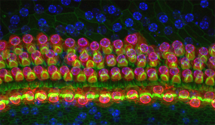
Outer plexiform layer of zebrafish retina |
|
Description |
Pedicles of 4 type of cones (UV Blue Green Red, bottom), 3 types of Horizontal Cell (H1 yellow, H2 and H3 orange), Many types of Bipolar cells (various colours, top). The dense neuropil of the OPL(near middle). The rod network and Mueller cells not shown. This is to illustrate the amazing complexity of the very first synaptic layer in the visual system which does a tremendous amount of information processing. |
|
Submission Date |
23.04.25 |
|
Name(s) of LEAD submitting author(s ) who must be members of the Anatomical Society and institution(s) |
Professor William A. Harris University of Cambridge |
|
Name(s) of Other submitting author(s ) and institution(s) |
None |
BEST IMAGE AWARD RUNNER- UP (ARTWORK CATEGORY)
|
Image Title |
|
|
Description
|
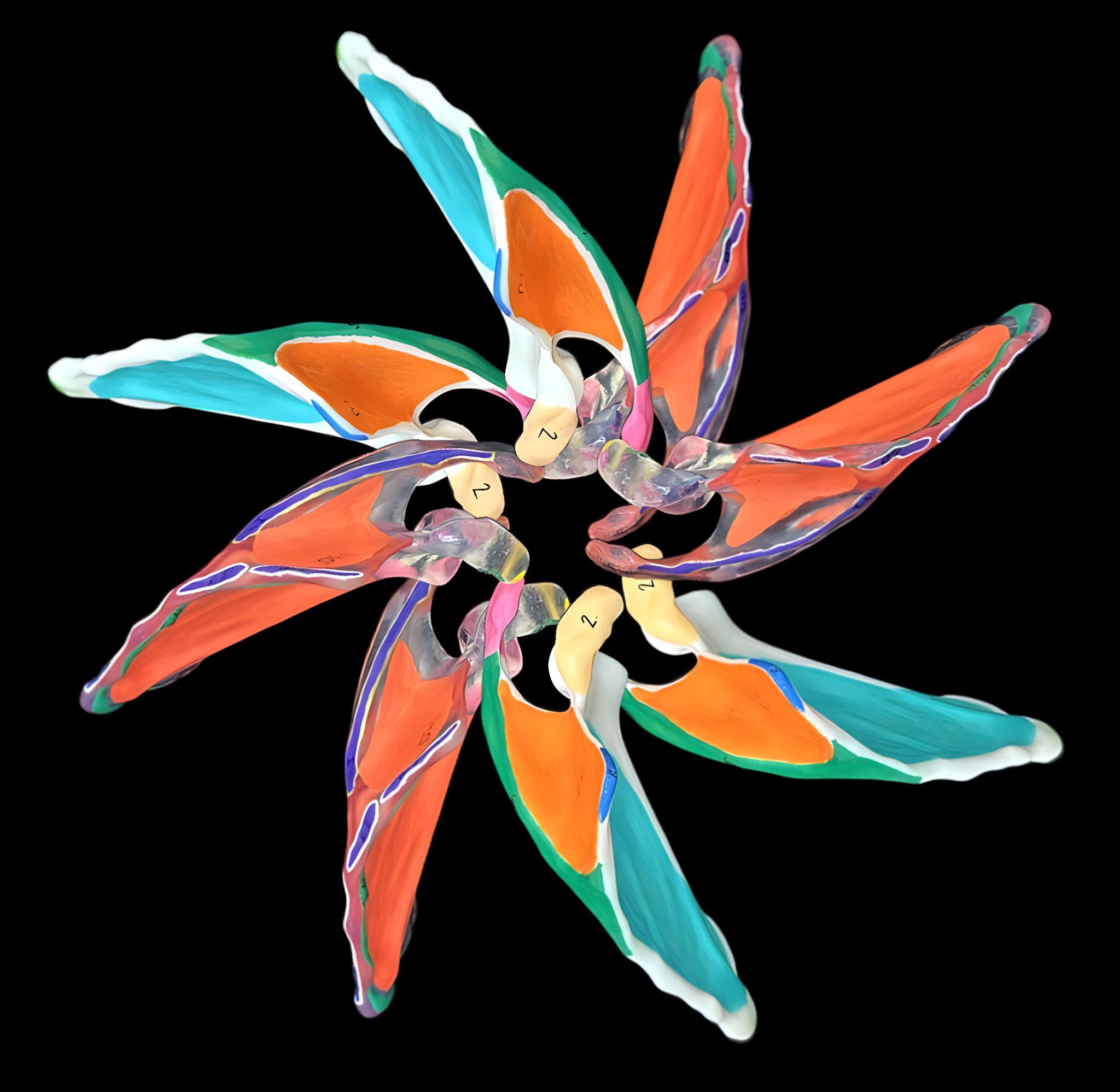
Scapulae were fabricated in white and clear resin from high-resolution scans to produce anatomically accurate models. These models were then hand-painted to highlight key skeletal landmarks and muscle origins and insertions to enhance their educational value. Two identical images of the models were overlayed to create the final floral patters. |
|
Submission Date |
29.05.25 |
|
Name(s) of LEAD submitting author(s ) who must be members of the Anatomical Society and institution(s) |
Dr Jennifer Z. Paxton University of Edinburgh |
|
Name(s) of Other submitting author(s ) and institution(s) |
Ms Iona Eldhose University of Edinburgh |
BEST IMAGE AWARD (SCIENCE CATEGORY)
|
Image Title |
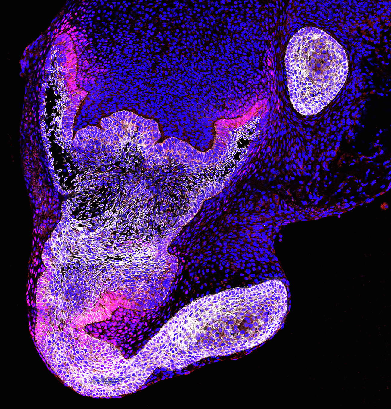
Devil Tooth Bud |
|
Description
|
Immunofluorescence of an explanted embryonic mouse molar treated with an inhibitor to Hedgehog signalling ex vivo. Dampening hedgehog signalling leads to ectopic tooth formation from the dental epithelium (Keratin14 in white) as shown by elevated Wnt signalling (Lef1 in red), highlighting the signals that control tooth number. Nuclei in blue. |
|
Submission Date |
30.05.25 |
|
Name(s) of LEAD submitting author(s ) who must be members of the Anatomical Society and institution(s) |
Professor Abigail Tucker |
|
Name(s) of Other submitting author(s ) and institution(s) |
Ms Litian Young King's College London |
BEST IMAGE AWARD RUNNER- UP (SCIENCE CATEGORY)
|
Image Title |
|
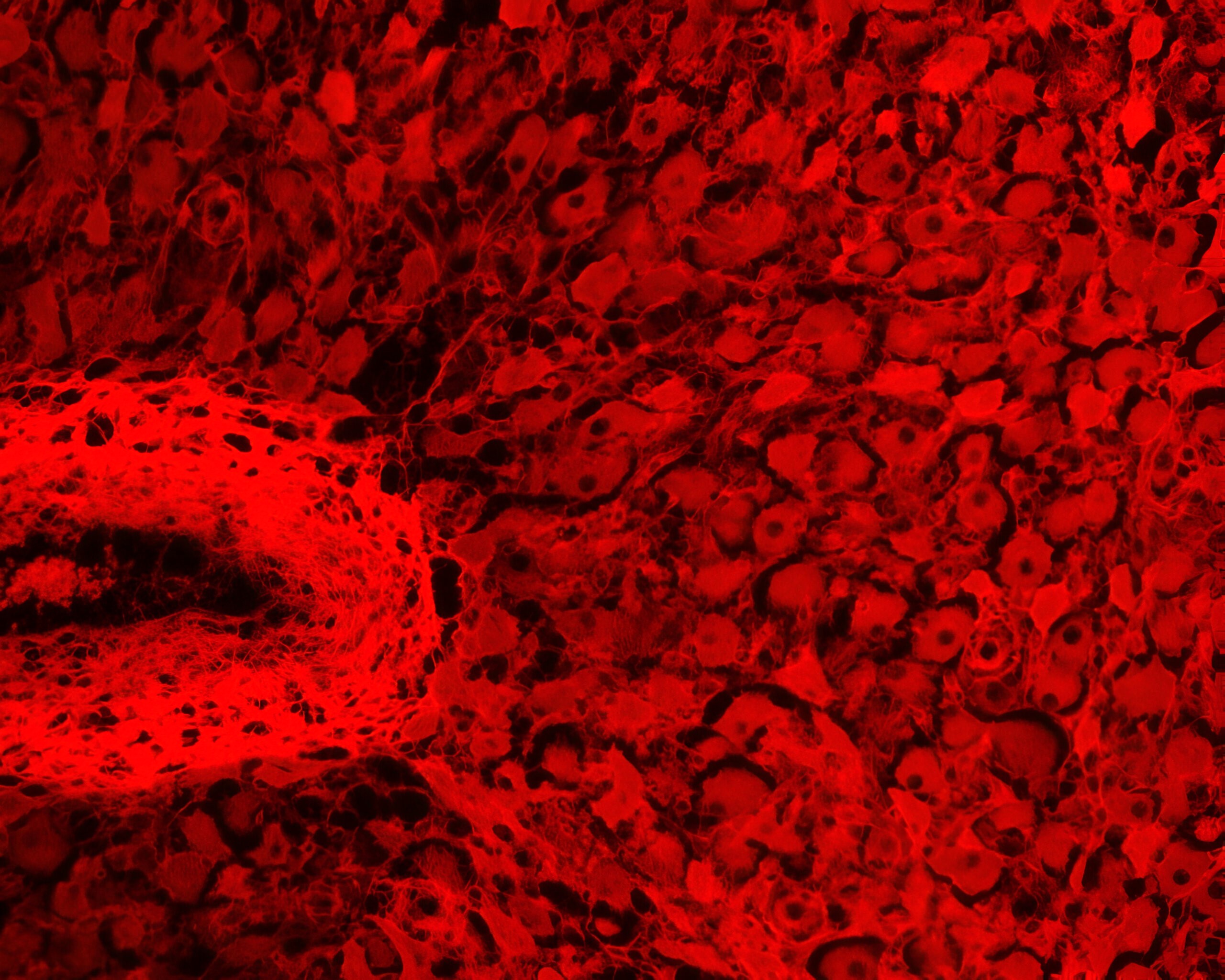
|
Description
|
Dorsal root ganglion (DRG) neurons extend processes across a microRNA-activated scaffold, showing guided neural outgrowth necessary for the repair of chronic wounds. The image captures the early stages of connection formation on the bio engineered microenvironment. The confocal image displays cell nuclei in blue and beta-tubulin III in white. |
|
Submission Date |
15.04.25 |
|
Name(s) of LEAD submitting author(s) who must be members of the Anatomical Society and institution(s) |
Mr Juan Carlos Palomeque |
|
Name(s) of Other submitting author(s) and institution(s) |
None |
|
Image Title |
|
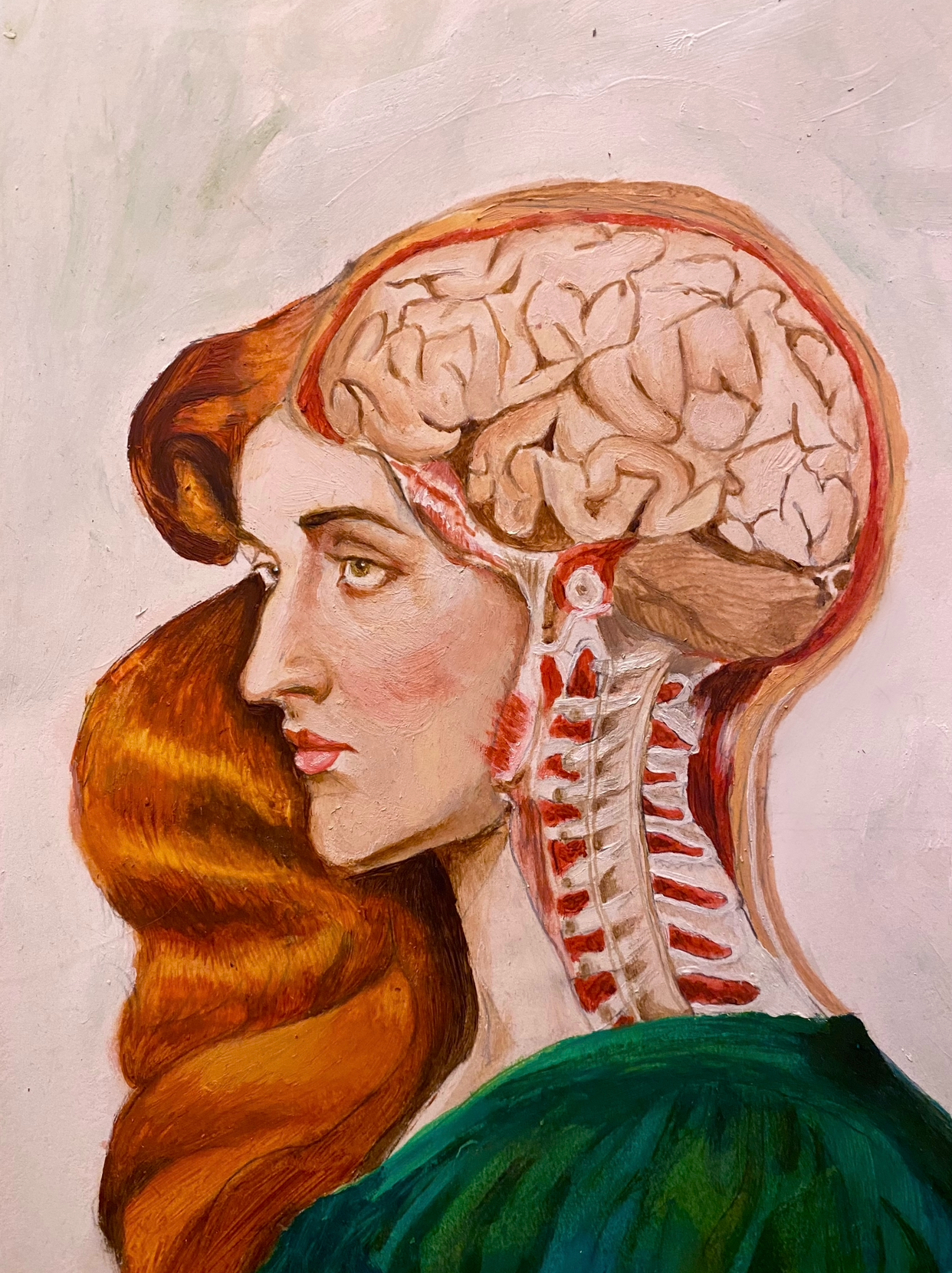
|
Description
|
A painting combining my two favourite elements: the aesthetics of neuroanatomy illustrations and the romantic style of Dante Gabriel Rossetti. Oil on canvas. |
|
Submission Date |
30.10.24 |
|
Name(s) of LEAD submitting author(s ) who must be members of the Anatomical Society and institution(s) |
Ms Diyya Ameen King’s College London |
|
Name(s) of Other submitting author(s ) and institution(s) |
None |
|
Image Title |
|
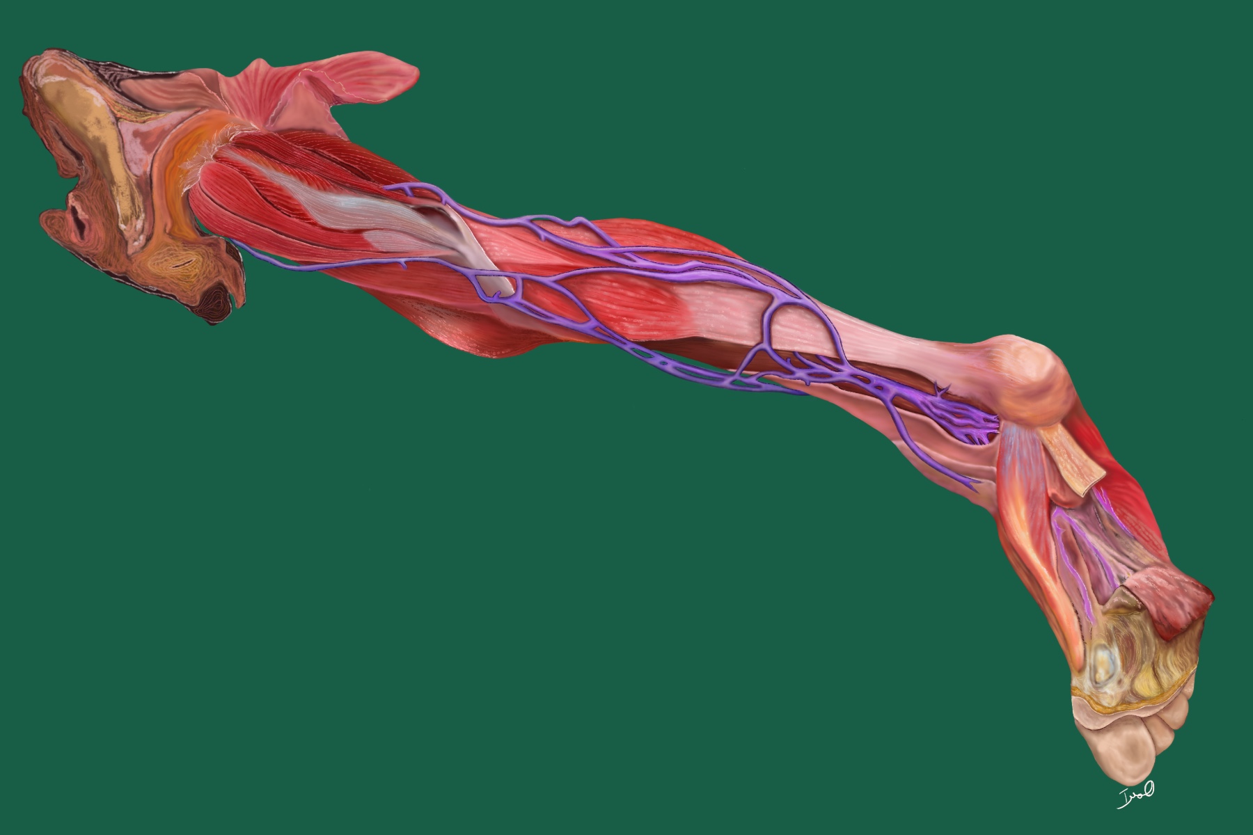
|
Description
|
Digital painting of a preserved wet specimen prepared to demonstrate the superficial muscles and vessels of the lower limb. This specimen highlights anatomical clarity in muscle orientation and vascular pathways, enhancing educational understanding of lower limb anatomy through precise visual representation. |
|
Submission Date |
31.10.24 |
|
Name(s) of LEAD submitting author(s ) who must be members of the Anatomical Society and institution(s) |
Mrs Humayra Bakar University of St Andrews |
|
Name(s) of Other submitting author(s ) and institution(s) |
None |
BEST IMAGE AWARD RUNNER- UP (ARTWORK CATEGORY)
|
Image Title |
|
|
Description
|
This digital artwork transforms the bronchial tree into a living grove. With bronchi resembling rugged trunks and alveoli forming a delicate canopy, each branch breathes life. Originating from a hand-drawn sketch, this piece was digitally enhanced, blending art and anatomy to capture the lungs' hidden vitality. |
|
Submission Date |
30.10.24 |
|
Name(s) of LEAD submitting author(s ) who must be members of the Anatomical Society and institution(s) |
Dr Noor Azzizah Omar Universiti Sains Islam Malaysia |
|
Name(s) of Other submitting author(s ) and institution(s) |
Ms Zulfa Mahiz Universiti Sains Islam Malaysia |
BEST IMAGE AWARD (SCIENCE CATEGORY)
|
Image Title |
|
|
Description
|
Double immunostaining revealed that the transcription factors FOXP2 and NR2F2 are expressed in the developing human thalamus at 10 post-conception weeks (pcw). Notably, NR2F2 is localized to the prethalamus,ganglionice eminence and hypothalamus , while FOXP2 is detected in postmitotic cells within the thalamus. |
|
Submission Date |
31.10.24 |
|
Name(s) of LEAD submitting author(s ) who must be members of the Anatomical Society and institution(s) |
Dr Maznah Abdullah Alhesain |
|
Name(s) of Other submitting author(s ) and institution(s) |
Dr Gavin Clowry Newcastle University |
BEST IMAGE AWARD RUNNER- UP (SCIENCE CATEGORY)
|
Image Title |
|
|
Description
|
Corpus luteum of pregnancy showing Small and Large Luteal Cells encapsulated (dark red outline) by collagen fibers; located adjacent to network of capillaries (for progesterone release), a bigger blood vessel (RBCs in lumen) surrounded by dense collagen fibers visualized by Fluorescence Microscopy stained by PicroSirius Red (20X; Fluorescent Collagen Stain). |
|
Submission Date |
11.10.24 |
|
Name(s) of LEAD submitting author(s) who must be members of the Anatomical Society and institution(s) |
Dr Kritima Kapoor |
|
Name(s) of Other submitting author(s) and institution(s) |
None |
BEST IMAGE PRIZE (ARTWORK CATEGORY)
|
Image Title |
|
|
Description
|
These 3D printed lung models were created from 3D scan data of a historic teaching model and painted in textbook and cadaveric colours. There is often a disconnect between what students see in textbooks vs the laboratory, therefore, these models aid to bridge the gap for students learning. |
|
Submission Date |
31.05.24 |
|
Name(s) of LEAD submitting author(s ) who must be members of the Anatomical Society and institution(s) |
Ms Victoria McCulloch, University of Edinburgh |
|
Name(s) of Other submitting author(s ) and institution(s) |
Ms Kilmeny Wong, University of Edinburgh |
RUNNER-UP BEST IMAGE PRIZE (ARTWORK CATEGORY)
|
Image Title |
|
|
Description
|
Neonatal mouse retinal ganglion cells undergo programmed cell death. Apoptotic cells on the edge of the growing vasculature can be labelled with specific markers such as Yo-PRO1 that fluoresce when binding to fragmenting DNA. Composition (arranged by blue-yellow, green-red colour opponency) inspired by Andy Warhol paintings. |
|
Submission Date |
31.05.24 |
|
Name(s) of LEAD submitting author(s ) who must be members of the Anatomical Society and institution(s) |
Professor Evelyne Sernagor, Newcastle University
|
|
Name(s) of Other submitting author(s ) and institution(s) |
Dr Michael Savage, Newcastle University Ms Cori Bertram, Newcastle University |
BEST IMAGE PRIZE (SCIENTIFIC CATEGORY)
|
Image Title |
|
|
Description
|
20-week-old male STR/Ort mouse knee, synchrotron scanned at Diamond Light Source (MG28353-1), in coronal plane. Pixels more than one standard deviation away from mean greyscale values have been highlighted in ImageJ, and false colours applied in photoshop tinting bone (blue), marrow (orange), cartilage (green) and calcified cartilage (purple). |
|
Submission Date |
15.05.24 |
|
Name(s) of LEAD submitting author(s ) who must be members of the Anatomical Society and institution(s) |
Dr Lucinda Evans, Royal Veterinary College, London and University of Brighton |
|
Name(s) of Other submitting author(s ) and institution(s) |
Mr Mark Hopkinson, Royal Veterinary College, London |
RUNNER-UP BEST IMAGE PRIZE (SCIENTIFIC CATEGORY)
|
Image Title |
|
|
Description
|
Mice embryo of E-13 day showing the expression of P53 in developing embryo. Embryos were retrieved from pregnant mice and immunoexpression of p53 was observed with respect to different stages of lungs development namely Pseudoglandular, Canalicular and Saccular. In this image, the lungs are in pseudoglandular stage of lungs development. |
|
Submission Date |
31.05.24 |
|
Name(s) of LEAD submitting author(s ) who must be members of the Anatomical Society and institution(s) |
Dr Amna Mughal, Dow University of Health Sciences, Pakistan |
|
Name(s) of Other submitting author(s ) and institution(s) |
Professor. Dr. Mushtaq Hussain, Dow University of Health Sciences, Pakistan |
|
mage Title |
Optic Chasm |
|
Description
|
Retinal axons in the optic chiasm and the optic tracts in an embryonic brain. The colours of the axons indicate regions of origins in the two retinas. Note that some axons cross and other don't, and the reordering of axons after the chiasm. |
|
Submission Date |
09.10.2023 |
|
Name(s) of LEAD submitting author(s ) who must be members of the Anatomical Society and institution(s) |
Professor William A. Harris University of Cambridge |
|
Name(s) of Other submitting author(s ) and institution(s) |
None |
RUNNER-UP BEST IMAGE PRIZE (ARTWORK CATEGORY)
|
Image Title |
Cephalopod Beak |
|
Description
|
Series of rendered images illustrating cephalopod beak opening (top to bottom) and closing (bottom to top) sequence. Obtained from CT reconstruction which was furthered rendered in Autodesk Fusion 360 |
|
Submission Date |
18.09.2023 |
|
Name(s) of LEAD submitting author(s ) who must be members of the Anatomical Society and institution(s) |
Dr Erwin Pauws University College London |
|
Name(s) of Other submitting author(s ) and institution(s) |
Mr Marius Didziokas University College London Dr Louise Souquet University College London |
BEST IMAGE PRIZE (SCIENTIFIC CATEGORY)
|
Image Title |
Invading Vasculature |
|
Description
|
During tooth development the vasculature (Red) surrounds the forming tooth and starts to invade the dental papilla in response to Wnt signalling (Green). Image shows a confocal 3D Z-stack of a murine embryonic tooth germ (E14.5). Lef1-positive cells (Green) and CD31-positive endothelial cells (Red) shown by immunofluorescence. Nuclei DAPI (Blue) |
|
Submission Date |
27.10.2023 |
|
Name(s) of LEAD submitting author(s ) who must be members of the Anatomical Society and institution(s) |
Professor Abigail Tucker King's College London |
|
Name(s) of Other submitting author(s ) and institution(s) |
Dr Hiba Asrar King's College London |
RUNNER-UP BEST IMAGE PRIZE (SCIENTIFIC CATEGORY)
|
Image Title |
Computational tissue differentiation |
|
Description
|
We developed a computational algorithm to stimulate the bone formation at sutures. This image briefly presents the procedures from initial skull reconstruction of a 3-month-age individual, to the development of baseline in silico, model, end with predicted model at 12-month-age by expansion of cranial cavities and tissue differentiations at sutures. |
|
Submission Date |
26.10.2023 |
|
Name(s) of LEAD submitting author(s ) who must be members of the Anatomical Society and institution(s) |
Mr Ce Liang University College London |
|
Name(s) of Other submitting author(s ) and institution(s) |
Mr Marius Didziokas University College London |
|
Image Title |
|
|
Description
|
This artwork displays the intricate details of the skull of a Whitetail Deer (Odocoileus virginianus), a native North American species of cultural and ecological significance. Created with charcoal and carbon dust on clay board, the image highlights osteology features unique to this important mammal. |
|
Submission Date |
16.05.23 |
|
Name(s) of LEAD submitting author(s ) who must be members of the Anatomical Society and institution(s) |
Professor Benjamin Crockett, North Central Michigan College, USA |
|
Name(s) of Other submitting author(s ) and institution(s) |
N/A |
RUNNER-UP BEST IMAGE PRIZE (ARTWORK CATEGORY)
|
Image Title |
|
|
Description
|
Painting on primed canvas with acrylics, inspired from CT angiography of head and neck; signifies the system of arteries supplying various parts of brain and spinal cord. The background symbolizes a black out in case of starvation. In a healthy individual, the nourishment provided is depicted as a glowing bulb. |
|
Submission Date |
30.05.23 |
|
Name(s) of LEAD submitting author(s ) who must be members of the Anatomical Society and institution(s) |
Professor Kalpana Ramachandran Sri Ramachandra Medical College & Research Institute(SRMC & RI), Sri Ramachandra Institute of Higher Education and Research (SRIHER), Chennai, Tamilnadu, India |
|
Name(s) of Other submitting author(s ) and institution(s) |
Ms Nivedha Saravanan Sri Ramachandra Medical College & Research Institute (SRMC & RI) Sri Ramachandra Institute of Higher Education and Research (SRIHER), Chennai, Tamilnadu, India.
|
BEST IMAGE PRIZE (SCIENTIFIC CATEGORY)
|
Image Title |
|
|
Description
|
A max projection from a 3-D Z stack of a pregnant ovine mammary gland, produced through confocal microscopy after CUBIC optical tissue clearing. Macrophages, stained with IBA-1 (magenta), are abundant in the gland and exhibit close associations with myoepithelial cells, stained with alpha-SMA (yellow). Nuclei are stained with DAPI (cyan). |
|
Submission Date |
26.05.23 |
|
Name(s) of LEAD submitting author(s ) who must be members of the Anatomical Society and institution(s) |
Mr Benjamin Davies, University of Cambridge |
|
Name(s) of Other submitting author(s ) and institution(s) |
Dr Kate Hughes, University of Cambridge |
RUNNER-UP BEST IMAGE PRIZE (SCIENTIFIC CATEGORY)
|
Image Title |
|
|
Description
|
A >100-day old patient-derived astrocyte grown on neurotrophic proteins is shown here using a technique known as stimulated emission depletion (STED) which allows for enhanced resolution, overcoming the diffraction limit of light. Here nanoscale differences in F-actin (blue) structures can be observed, with enhanced micro-processes extension on the trophic proteins. |
|
Submission Date |
11.05.23 |
|
Name(s) of LEAD submitting author(s ) who must be members of the Anatomical Society and institution(s) |
Mr Cian O’Connor, Tissue Engineering Research Group, Dept of Anatomy & Regenerative Medicine, Royal College of Surgeons in Ireland, Dublin, Ireland |
|
Name(s) of Other submitting author(s ) and institution(s) |
Mr Massimiliano Garre, Super Resolution Imaging Consortium, Royal College of Surgeons in Ireland, Dublin, Ireland Ms Rena Mullally, Tissue Engineering Research Group, Dept of Anatomy & Regenerative Medicine, Royal College of Surgeons in Ireland, Dublin, Ireland |
|
Image Title |
|
|
Description
|
A 3D maximum projection of a whole mount chicken embryo at stage HH11 constructed from a confocal stack. Hybridization chain reaction shows Uncx4.1 (green) and EphA4 (magenta) alongside DAPI (cyan). This reveals the rostral (EphA4) and caudal (Uncx4.1) identity of cells in newly forming somites. |
|
Submission Date |
25/10/2022 |
|
Name(s) of LEAD submitting author(s) who must be members of the Anatomical Society and institution(s) |
Mr Adam Moverley University College London |
|
Name(s) of Other submitting author(s) and institution(s) |
Professor Claudio Stern University College, London |
|
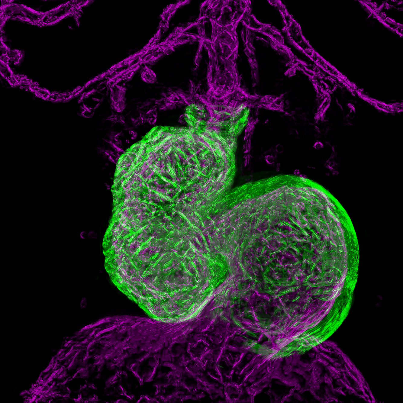
Image Title |
|
|
Description
|
Confocal images (40X objective) of the structure and vasculature of the heart from a 18-day old mouse. A tranverse section of the heart (thickness 8µm) is immunostained to visualise cell membranes (red), endothelial cells (green, here seen as yellow since green overlaps with red) and cell nuclei (blue). |
|
Submission Date |
27/10/22 |
|
Name(s) of LEAD submitting author(s) who must be members of the Anatomical Society and institution(s) |
Ms Nithya N. Nair University of Edinburgh |
|
Name(s) of Other submitting author(s) and institution(s) |
N/A |
JOINT RUNNER-UP BEST IMAGE PRIZE
|
Image Title |
|
|
Description
|
A 3D reconstruction and cross section of a mutant mouse heart collected at embryonic day (E) 15.5. Sections are produced using High Resolution Episcopic Microscopy (HREM) and processed by multiple image editing software that finally allows for accurate phenotypic analysis. This cross section shows a perimembranous ventricular septal defect. |
|
Submission Date |
28/10/22 |
|
Name(s) of LEAD submitting author(s) who must be members of the Anatomical Society and institution(s) |
Ms Aseel Abbad University of Nottingham |
|
Name(s) of Other submitting author(s) and institution(s) |
Dr Siobhan Loughna University of Nottingham Professor David Brook University of Nottingham |
|
Image Title |
|
|
Description
|
The skull of a new fossil salamander from the Middle Jurassic of Scotland in front view. The image was created following X-ray microCT scan of fossil blocks, segmentation of individual bones in Amira 6.0, and reorientation of individual bones in Blender 2.9. The eyes are to indicate orbit location. |
|
Submission Date |
31/10/22 |
|
Name(s) of LEAD submitting author(s) who must be members of the Anatomical Society and institution(s) |
Dr Marc Jones University College London |
|
Name(s) of Other submitting author(s) and institution(s) |
Professor Roger Benson University of Oxford Professor Susan Evans University College London
|
|
Image Title |
|
|
Description
|
SiR-actin labelling in stereocilial bundles from live auditory hair cells. |
|
Submission Date |
22/09/22 |
|
Name(s) of LEAD submitting author(s) who must be members of the Anatomical Society and institution(s) |
Dr Zoe F. Mann King’s College London |
|
Name(s) of Other submitting author(s) and institution(s) |
Dr James O’Sullivan King’s College London |
|
Image Title |
|
|
Description
|
Breast cancer cell engaging in cross-talk with collagen fibrils in the extracellular matrix (ECM). This image taken by SEM of breast cancer cells cultured on collagen-based scaffolds, captures the extraordinary interactions between a breast cancer cell and its surrounding ECM which plays a major role in cancer progression. |
|
Submission Date |
25.05.22 |
|
Name(s) of LEAD submitting author(s ) who must be members of the Anatomical Society and institution(s) |
Ms Elizabeth Sainsbury, Royal College of Surgeons in Ireland |
|
Name(s) of Other submitting author(s ) and institution(s) |
N/A |
JOINT RUNNER-UP PRIZE
|
Image Title |
|
|
Description
|
This digital artwork was created as a part of a self-directed project. Each image depicts the gross and microanatomy of the kidneys. I tried to create a unique style by combining traditional line drawing techniques with a hint of traditional Asian artwork. |
|
Submission Date |
30.05.22 |
|
Name(s) of LEAD submitting author(s) who must be members of the Anatomical Society and institution(s) |
Ms Daheen Lee |
|
Name(s) of Other submitting author(s) and institution(s) |
N/A |
|
Image Title |
|
|
Description
|
Murine Eustachian Tube. Basal epithelial stem cells (Keratine 5 in green) give rise to two types of cells responsible for pathogens clearance from the middle ear. Goblets cells (plunc1 in white) produce mucines that are pushed towards the throat by Ciliated cells (acetylated tubuline in red). Nuclei in Blue (DAPI). |
|
Submission Date |
20.05.22 |
|
Name(s) of LEAD submitting author(s) who must be members of the Anatomical Society and institution(s) |
Dr Juan Manuel Fons Romero |
|
Name(s) of Other submitting author(s) and institution(s) |
N/A |
|
Image Title |
|
|
Description
|
The white lightning bolt-like strands you see connecting and surrounding collagen IV-expressing cells (blue) are depositions of an extracellular protein, fibronectin (white). Here we see its deposition by human meningeal fibroblasts as they are grown on top of human spinal cord astrocytes. Red= cell nuclei. |
|
Submission Date |
31.05.22 |
|
Name(s) of LEAD submitting author(s) who must be members of the Anatomical Society and institution(s) |
Dr Martyna Stasiewicz |
|
Name(s) of Other submitting author(s) and institution(s) |
N/A |
|
Image Title |
|
|
Description
|
Graphite pencil and watercolour of a horse femur |
|
Submission Date |
07/10/21 |
|
Name(s) of LEAD submitting author(s) who must be members of the Anatomical Society and institution(s) |
Dr Fay Penrose, University of Liverpool, UK |
|
Name(s) of Other submitting author(s) and institution(s) |
N/A |
RUNNER-UP PRIZE
|
Image Title |
|
|
Description
|
‘The Anatomy Lesson of Dr. Nicolaes Tulp’ (Rembrandt 1632) shows students studying cadaveric anatomy in the laboratory. Inspired by this painting’s composition, I created the ‘COVID-safe Anatomy Lesson’ in this acrylic on A3 canvas piece. My apologies to Rembrandt. |
|
Submission Date |
30/09/21 |
|
Name(s) of LEAD submitting author(s) who must be members of the Anatomical Society and institution(s) |
Professor Ian Johnson, Macquarie University,Sydney, Australia |
|
Name(s) of Other submitting author(s) and institution(s) |
N/A |
|
Image Title |
|
|
Description
|
Pseudo-colorized scanning electron micrograph of the in vitro mineralised jellyfish collagen scaffold (Jellagen®Ltd). Owing to the highly porous structure of the scaffold, collagen fibres (PINK and GREEN) are randomly tangled in a flower-like shape, with mineral deposition (yellow) observed on the surface for enhanced osseointegration and new bone formation. |
|
Submission Date |
24/05/2021 |
|
Name(s) of LEAD submitting author(s ) who must be members of the Anatomical Society and institution(s) |
Miss Patricia Medesan, Doctoral Fellow in Anatomical/Biomedical Sciences, Paxton Laboratory, Edinburgh Medical School, UK |
|
Name(s) of Other submitting author(s ) and institution(s) |
Dr Jennifer Z. Paxton, Senior Lecturer in Anatomy, Centre for Discovery Brain Sciences, Edinburgh Medical School, UK Professor Andrew Mearns-Spragg, Jellagen® Ltd, Cardiff, UK |
RUNNER-UP PRIZE
|
Image Title |
|
|
Description |
As an artist with a fascination for Anatomy, I am privileged to have the opportunity to carry out cadaveric dissections. This oil painting (120cm x 120cm) is my subjective response to that necessarily objective experience. The intention is to challenge what I see as the tenuous art - science distinction. |
|
Submission Date |
20/05/21 |
|
Name(s) of LEAD submitting author(s ) who must be members of the Anatomical Society and institution(s) |
Dr Jac Saorsa |
|
Name(s) of Other submitting author(s ) and institution(s) |
None |
|
FIRST PRIZE |
|
|
Image Title |
|
|
Description
|
Illustration of the right eye showing the ophthalmic and maxillary divisions of the trigeminal nerve. Drawn by hand using pencil with computer colour rendering. Completed for research on the disease of syphilis. |
|
Submission Date |
31/10/20 |
|
Name(s) of LEAD submitting author(s ) who must be members of the Anatomical Society and institution(s) |
Ms Lydia Carline |
|
Name(s) of Other submitting author(s ) and institution(s) |
N/A |
|
RUNNER-UP BEST IMAGE PRIZE |
|
|
Image Title |
|
|
Description |
A maximum image projection of a differentiating induced pluripotent stem cell derived neurosphere cultured in a 3D porous, soft hyaluronic acid tissue scaffold in vitro. Astrocytes (pink) coalesce at the core driving intrinsic signalling while differentiating neurons (yellow) are found at the periphery. Imaged using a Zeiss Examiner.Z1 confocal microscope. |
|
Submission Date |
29/10/2020 |
|
Name(s) of LEAD submitting author(s ) who must be members of the Anatomical Society and institution(s) |
Dr Adrian Dervan - Royal College of Surgeons in Ireland & Advanced Materials and Bioengineering Research Centre (AMBER) |
|
Name(s) of Other submitting author(s ) and institution(s) |
Mr Cian O’ Connor – Royal College of Surgeons in Ireland & Advanced Materials and Bioengineering Research Centre (AMBER) Professor Fergal O’Brien - Royal College of Surgeons in Ireland & Advanced Materials and Bioengineering Research Centre (AMBER) |
|
FIRST PRIZE |
|
|
Image Title |
|
|
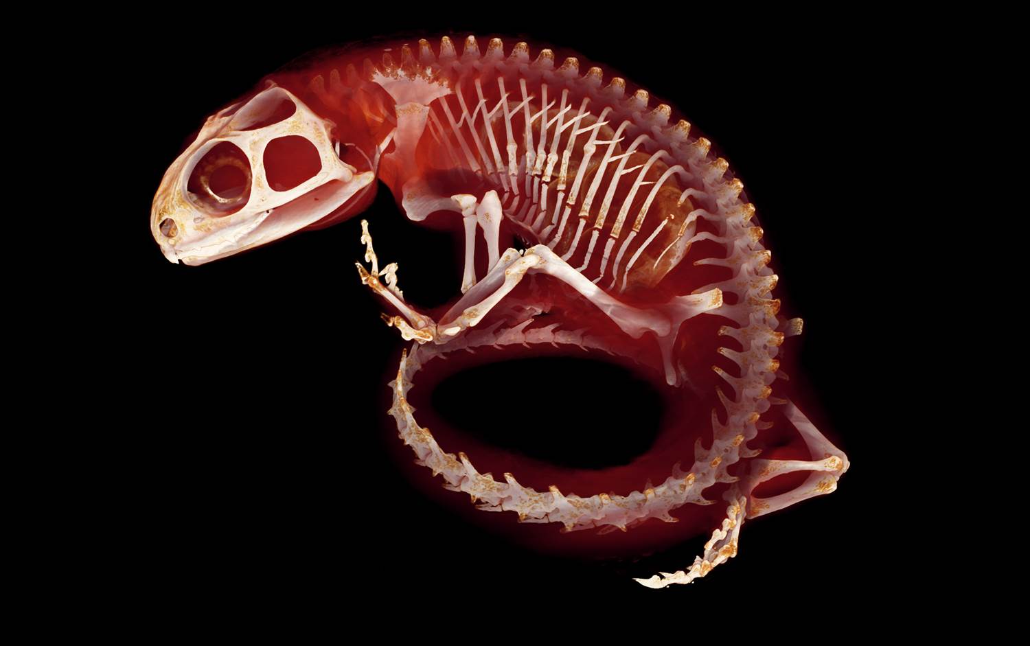
Description
|
In this skeletal preparation of a little skate (Leucoraja erinacea) hatchling, cartilage and mineralised tissues are stained with Alcian Blue and Alizarin Red, respectively. Cartilaginous fishes, like the skate, do not form bone, but instead reinforce their skeleton with tiny superficial tiles of calcified cartilage called “tesserae”. |
|
Submission Date |
30 May 2020 |
|
Name(s) of LEAD submitting author(s ) who must be members of the Anatomical Society and institution(s) |
Andrew Gillis |
|
Name(s) of Other submitting author(s ) and institution(s) |
N/A |
|
RUNNER-UP PRIZE |
|
|
Image Title |
|
|
Description
|
Confocal image of the root of a tooth showing in green the expression of the putative stem cell marker Thy1. The image has been mirrored and flipped to give the impression of a kaleidoscope. |
|
Submission Date |
27/05/2020 |
|
Name(s) of LEAD submitting author(s ) who must be members of the Anatomical Society and institution(s) |
Abigail Tucker |
|
Name(s) of Other submitting author(s ) and institution(s) |
Rupali Lav |
|
FIRST PRIZE |
|
|
Image Title |
|
|
Description
|
Live light sheet images of the heart and connecting vasculature of a 3-day old zebrafish embryo. Tissue-specific transgenic lines allow visualisation of actin in the myocardial (green) and endocardial (magenta) cells of the heart, providing highly detailed 3D reconstructions of cardiac morphogenesis. |
|
Submission Date |
29.10.19 |
|
Name(s) of LEAD submitting author(s ) who must be members of the Anatomical Society and institution(s) |
Dr Emily Noël, University of Sheffield |
|
Name(s) of Other submitting author(s ) and institution(s) |
Ms Juliana Sanchez Posada |
|
JOINT RUNNER-UP |
|
|
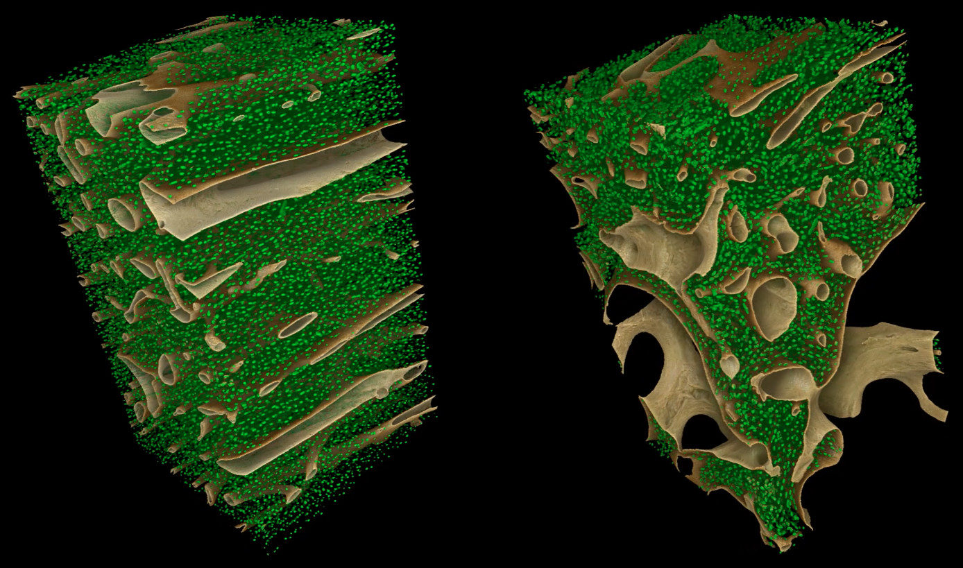
Image Title |
|
|
Description
|
High-resolution µCT data was collected at the Diamond Light Source synchrotron to visualise the internal structure of human orthotopic (left) and heterotopic (right) bone. This 3D volumetric render illustrates the morphological comparability of osteocyte lacunae (in green) across bone types. Bone surfaces (including Haversian canal walls) are shown in brown. |
|
Submission Date |
31st October, 2019 |
|
Name(s) of LEAD submitting author (s) who must be members of the Anatomical Society and institution(s) |
Dr Crispin Wiles, University of Warwick |
|
Name(s) of Other submitting author (s) and Institution(s) |
None |
|
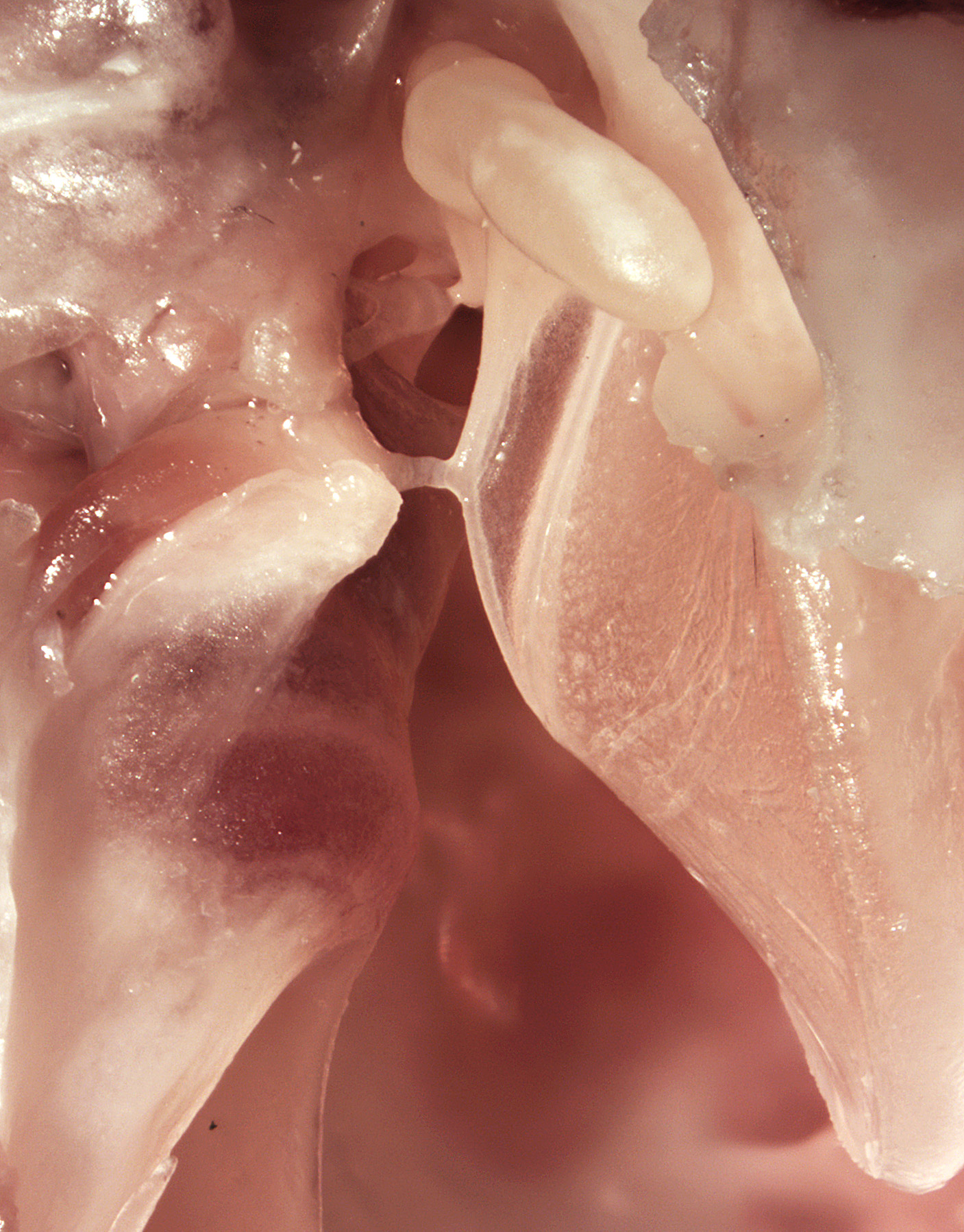
JOINT RUNNER-UP |
|
|
Image Title
|
|
|
Description
|
Composite photomicrograph of the left middle ear apparatus of a chinchilla. Careful dissection has revealed the tensor tympani muscle (centre left): its tendon crosses the middle ear cavity to insert on the manubrium of the malleus. The head of the stapes is visible in the background.
|
|
Submission Date |
31.10.19
|
|
Name(s) of LEAD submitting author (s) who must be members of the Anatomical Society and institution(s) |
Dr Matthew J. Mason University of Cambridge, Department of Physiology, Development & Neuroscience
|
|
Name(s) of Other submitting author (s) and institution(s) |
None |

|
Image Title (3 words max.) |
Cerebellar slice culture |
|
Description (50 words max.) |
This confocal image depicts a mouse cerebellar slice culture immunostained with the neuronal marker Calbindin (red) and myelin (white). Image was captured using a Zeiss LSM710 confocal microscope (Zeiss, Germany) and 10x objective.
|
|
Submission Date |
23/05/2019 |
|
Name(s) of LEAD submitting author(s ) who must be members of the Anatomical Society and institution(s)
|
Dr Francesca Pieropan
University of Portsmouth |
|
Name(s) of Other submitting author(s ) and institution(s) |
Dr Andrea D. Rivera
University of Portsmouth |
RUNNER-UP

|
Image Title (3 words max.) |
Warhol retinal clusters
|
|
Description (50 words max.) |
We recently discovered transient cholinergic cell clusters forming an annulus around the optic disc in the neonatal mouse retina. These images illustrate the clusters (ChAT immunostaining) in a retinal wholemount (postnatal day 5) with ganglion cells (RBPMS immunostaining) in the background in various colour combinations inspired by Andy Warhol’s paintings. |
|
Submission Date |
31 May 2019 |
|
Name(s) of LEAD submitting author(s ) who must be members of the Anatomical Society and institution(s) |
Professor Evelyne Sernagor |
|
Name(s) of Other submitting author(s ) and institution(s) |
Jean de Montigny Dr Vidhyasankar Krishnamoorthy Institute of Neuroscience, Newcastle University |
FIRST PRIZE

|
Image Title
|
Reconstructing Gigantic Dormouse |
|
Description
|
A 3D-model of one of the best-preserved skulls of the gigantic extinct Sicilian dormouse, Leithia melitensis, is created using photogrammetry. The missing cranial features, including the nasal bone, the incisors and the zygomatic arch, are reconstructed using a warped microCT of a smaller but closely related extant dormouse Eliomys quercinus from Sicily. |
|
Submission Date |
11.10.18 |
|
Name(s) of LEAD submitting author(s ) who must be members of the Anatomical Society and institution(s) |
Mr Jesse James Hennekam, |
|
Name(s) of Other submitting author(s ) and institution(s) |
None |
JOINT RUNNER-UP

|
Image Title |
Lonely Cortical Neuron |
|
Description |
A mouse pyramidal neuron marked by expression of tomato-DsRed. The neurons, detectedfollowingan elegant lineagetracing paradigm,reside in LayersII/III amongst existing cortical neurons, marked by NeuN immunolabelling, and Hoescht to detect nuclei. Imaged as a Z stack with a Zeiss Axiogimager M2 withapotome attachment. |
|
Submission Date |
12.10.18 |
|
Name(s) of LEAD submitting author(s ) who must be members of the Anatomical Society and institution(s) |
Miss Hannah Louisa Felstead (School of Biological Sciences, University of East Anglia) |
|
Name(s) of Other submitting author(s ) and institution(s) |
Dr. Mohammad K. Hajihosseini (School of Biological Sciences, University of East Anglia) |

|
Image Title |
Bearded seal maxilloturbinates
|
|
Description |
Micro-CT image of the nasal cavity of a bearded seal, showing a superimposed section of its extraordinarily complex maxilloturbinate bones. Owing to inevitably limited scan resolution, isolating the extremely thin turbinates as evenly as this, across the whole cavity, required considerable image processing and manual work. |
|
Submission Date |
2.10.18 |
|
Name(s) of LEAD submitting author(s ) who must be members of the Anatomical Society and institution(s) |
Dr Matthew James Mason, University of Cambridge
|
|
Name(s) of Other submitting author(s ) and institution(s) |
1) Léa Wenger, University of Cambridge 2) Øyvind Hammer, Natural History Museum, University of Oslo, Norway 3) Arnoldus Schytte Blix, Department of Arctic and Marine Biology, University of Tromsø, Tromsø, Norway |
|
FIRST PRIZE Image Title: Retina Alpha Other Submitting Author (s):Dr Gerrit Hilgen and Ms Viktoriia Kartysh, Institute of Neuroscience, Newcastle University. Submitted on: 31 May 2018. |
|
RUNNER-UP Image Title: The Arbor Vitae of the murine cerebellum Submitted on: 31 May 2018. |
|
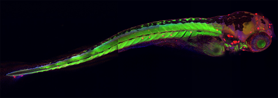
FIRST PRIZE |
|
|
Image Title: |
|
|
Description: |
5-day-old zebrafish larva stained with DAPI to label nuclei (blue), phalloidin to visualize F-actin (green) and Myosin7a to stain mechanosensory hair cells (red). Zebrafish sense water motion through hair cells (red) in neuromasts in the lateral line along the body and all over the head. |
|
Lead Submitting Author |
Professor Andrea Streit, King’s College London
|
|
Other Submitting Author: |
Ms Alice Gervasoni, King's College London |
|
Submitted on: |
04 October 2017 |
|
RUNNER-UP PRIZE |
|
|
Image Title: |
|
|
Description: |
P8 mouse cochlea stained with DAPI to label cell nuclei (blue), phalloidin (green) to visualize F-actin and Myosin7a (red) to stain mechanosensory hair cells. Mammals detect sounds through an array of sensory receptors called hair cells (red) characterized by stereocilia (green) on their apical surface. |
|
Lead Submitting Author: |
Professor Andrea Streit, King’s College London |
|
Other Submitting Author: |
Ms Alice Gervasoni, King's College London |
|
Submitted on: |
04 October 2017 |
|
FIRST PRIZE |
|
|
Image Title: |
|
|
Description: |
This is a preserved tuatara that has been imaged using X-ray microtomography. In addition to the skeleton, scleral ossicles, tiny sesamoid bones, and eggs within the abdomen can be seen. This was one of several rare and historically valuable tuatara specimens to be scanned and made freely available athttps://osf.io/bds35/.
|
|
Lead Submitting Author |
Sophie Regnault (Structure and Motion Lab, Royal Veterinary College)
|
|
Submitted on: |
1 February 2017
|
|
RUNNER-UP PRIZE |
|
|
Image Title: |
|
|
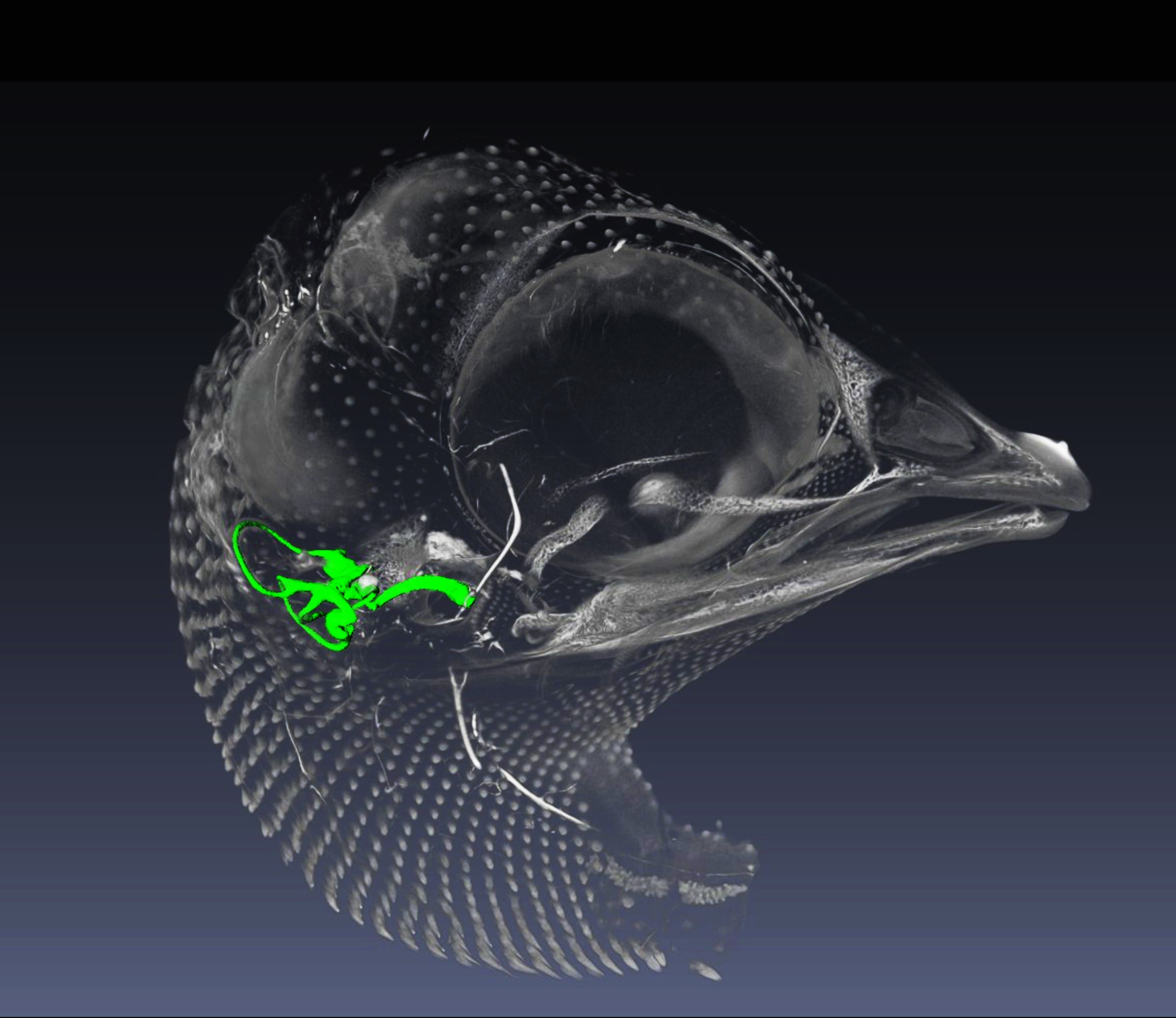
Description: |
A micro-CT reconstructed image of a 10-day-old chick embryo with the inner ear structures. The semicircular canals, vestibule and cochlea are highlighted in green
|
|
Lead Submitting Author:
|
Nobue Itasaki, University of Bristol |
|
Submitted on: |
21 April 2017 |
|
FIRST PRIZE |
|
|
Image Title: |
|
|
Description: |
The image depicts Myelin (Cyan) and Oligodendrocytes Precursor Cells (OPCs, Red) jn Corpus Callosum of a mouse brain aged Postnatal day (P) 8. Images were captured using a Zeiss LSM710 confocal microscope (Zeiss, Germany) and Plan-NEOFLUAR 20 objectives with a numerical aperture of 0.50. |
|
Lead Submitting Author |
Andrea D. Rivera,University of Portsmouth
|
|
Submitted on: |
29 October 2016
|
|
RUNNER-UP |
|
|
Image Title: |
Image Title:Human Fetal Forebrain |
|
Description: |
Description:Sagittal section of 8 post-conception week human early fetal brain (obtained fromwww.hdbr.org) stained by double label immunofluorescence for the cell division marker KI67 (green) andthe transcription factor COUPTFII (red). The combination highlights (yellow) regionalisation of COUPTFII-expressing dividing cells to the caudal ganglionic eminence. |
|
Lead Submitting Author: |
Ayman Alzu’bi, Newcastle University
|
|
Submitted on: |
8 September 2016 |
|
AS BEST IMAGE AWARD 2016 (31.05.16) |
|
|
Image Title: |
|
|
Description: |
3D anatomy of an embryonic day (E) 16.5 mouse forelimb produced by optical projection tomography (OPT). The merged image shows muscle (red) stained with an antibody against muscle myosin (My32). Tendons (green) are labelled using a ScxGFP reporter. Autofluorescence (grey) provides an outline of all limb structures. |
|
Submitted by Lead Author (Member AS):
|
Professor Malcolm Logan |
|
Submitted by Other Author:
|
Dr Susan Miller Randall Division, King’s College London |
|
Submitted on: |
26 April 2016 |
|
AS RUNNER-UP BEST IMAGE AWARD 2016 (31.05.16) |
|
|
Image Title: |
|
|
Description: |
This is an Old World fruit bat embryo collected in 1972 at Ife University, Nigeria. The specimen has recently been processed for histological sectioning to investigate the process of tooth replacement, showing an example of archival tissue being used in modern day science. Image obtained with a Leica MZFLIII stereoscope. |
|
Submitted by Lead Author (Member AS) |
Dr Neal Anthwal King’s College, London
|
|
Submitted byOther Author(s) |
Ms Elena Popa |
|
Submitted on: |
31 May 2016 |
S BEST IMAGE AWARD 2015 (31.10.15) AWARDED TO DR SUSAN CHAPMAN BY THE AS WEBSITE OFFICER

Image Title: Full Frontal Face
Description: A frontal section of an embryonic day 6 chicken head, showing the inner and middle ear, 2nd pharyngeal arches and tongue. Peanut Agglutinin Lectin (red), Sambucus nigra (green) and Wheat Germ Agglutinin (purple) fluorescently label neural tube, cartilage matrix, endothelial cells and epithelia. Lectin staining differentiates between cartilage elements.
Submitted by: Dr Susan C. Chapman, Clemson University
Submitted on: 13 October 2015
AS RUNNER-UP BEST IMAGE AWARD 2015 (31.10.15) AWARDED TO STEPHANE BERNEAU BY THE AS WEBSITE OFFICER

Image Title: In-vitro embryo invasion
Description: Fluorescent image of an advanced in vitro mouse embryo attachment site exhibiting embryo outgrowth into human endometrial Ishikawa cells to detect OPN receptor, CD44, in green.Endometrial CD44 plays a role in embryo attachment and its localisation remains unchanged at embryo invasion site.Blue: Cell nuclei staining. Red: Actin cytoskeleton.
Submitted by: Stéphane Berneau, University of Manchester, Institute of Human Development, Maternal and Fetal Health Research Group
Submitted on: 31 October 2015
AS BEST IMAGE AWARD 2015 (31.05.15)
Image Title: L1 vertebral trabeculae
Description: A sphere digitally cut from a 2.5-year old human L1 vertebral centrum and coloured to highlight variation in trabecular thickness. It was created from microCT data using VGStudio and Drishti, for an ongoing project comparing the ontogeny of vertebral trabeculae in modern humans and Neanderthals.
Submitted by: Crispin Wiles
Submitted on: 31 May 2015
AS RUNNER-UP BEST IMAGE AWARD 2015 (31.05.15)
Image Title: Peeling octopus sucker
Description: A sucker of Octopus vulgaris analysed by scanning electron microscopy (SEM) was captured shedding its skin. A Cambridge Stereoscan360 (Image Access, V.3) was used for acquisition; Adobe Photoshop for pseudocolouring (sucker, green; shedding skin, yellow). This micrograph of the normal shedding process reveals the sophisticated nature of the octopus skin’s interdigitations (comparable to rete ridges in human epidermis).
Submitted by: Tanya Shaw
Submitted on: 31 May 2015
AS BEST IMAGE AWARD 2014 (31.10.14)
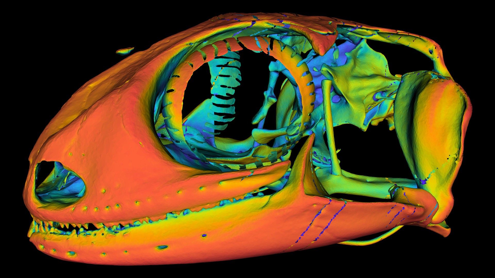
Image Title: Gecko ‘Chromocranium’
Description: 3D rendering of a gecko skull (Nephrurus levis) from micro-computed tomography (21 um/voxel). Colour corresponds to ambient occlusion (i.e. red surfaces are most visible, blue surfaces least visible). Steep colour gradients highlight foramina. Hidden structures (e.g. bony labyrinth, nasal cavity) can be extracted automatically based on their high occlusion.
Submitted by: Mr James Turbett
Submitted on: 30 October 2014
AS RUNNER UP BEST IMAGE AWARD 2014 (31.10.14)
Image Title: The Mummy's Return
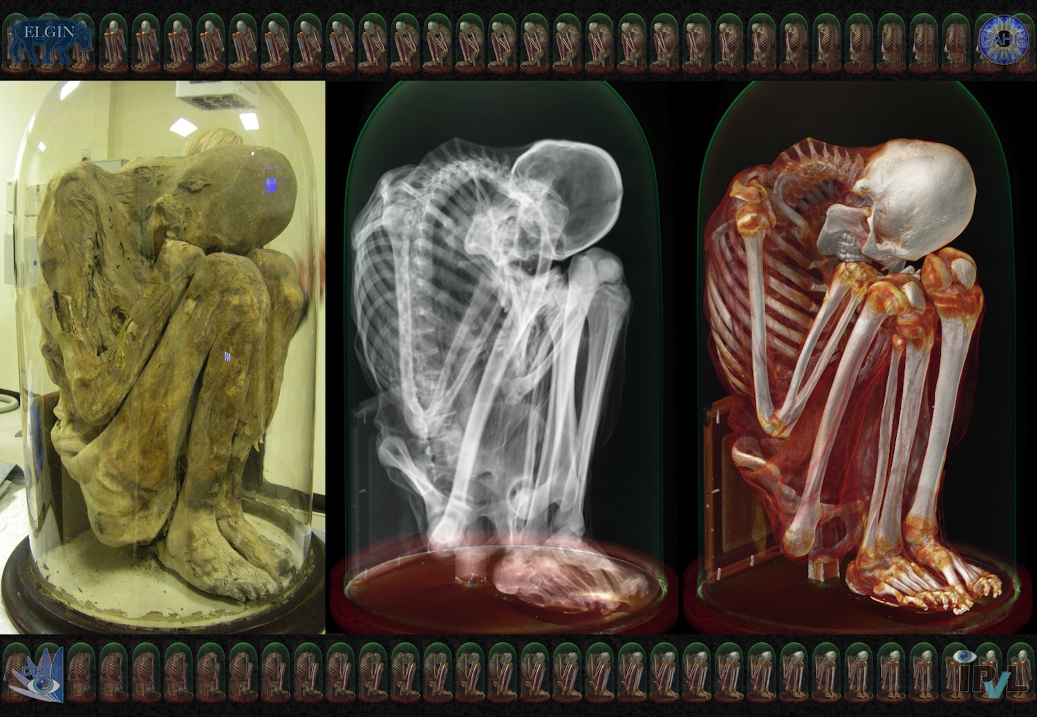
Description: A 15th century Peruvian mummy in a sealed glass-bell underwent forensic "phenotyping". Central to this study, a set of CT
scans led to subsequent advanced analysis, 3D-renderings and full body/face reconstructions, which together provided critical information
about the gender, age, condition and other aspects of the person prior to her death.
Submitted by: Professor Luc Bidaut
Submitted on: 28 October 2014
AS BEST IMAGE AWARD 2014 (deadline 31.05.14)
Image Title: Spinal astrocytes
Description: Image shows the morphology of rodent spinal astrocytes, which play an important role in numerous functions in the central nervous system. Vimentin (green) demarcates the cytoskeletal structure, illustrating the extensive processes of these cells and dapi (blue) shows the nucleus. The image was captured using a Zeiss LSM710 Confocal microscope.
Submitted by: Dr Ada Delaney
Submitted on: 14th May 2014
AS RUNNER UP BEST IMAGE AWARD 2014 (deadline 31.05.14)
Description: Mouse embryo at embryonic stage E12.5 labeled with DiI, a plasma membrane marker which was incorporated in the entire epithelial surface. By tracing cell movements, key roles of tissue interactions in many aspects of embryonic morphogenesis can be investigated, contributing to the developmental biology knowledge.
Submitted by: Ms Lemonia Chatzeli, Ms Tathyane Harumi Nakajima Teshima and Dr Marcia Gaete
Submitted: 15th May 2014
FIRST PRIZE
Description: Enteric neurospheres, generated from Wnt-1Cre;R26YFP mouse gut. Wnt-1 positive neural crest cells express YFP (green). The neurospheres are stained with dapi (blue) and immunolabelled with (from top to bottom in red) p75, TuJ1, GFAP, and Sox10. These immunomarkers confirm that gut neurospheres contain neural crest-derived cells, neurons, glia and ENS stem cells.
Submitted by: Dr Alan Burns and Dr Ellen Binder.
Submitted on: 25 October 2013
JOINT RUNNER-UP PRIZE
Image Title: Enhanced Tractography
Description: Axial diffusion-weighted MRI tractography image of living human brain. Nerve fibre colour represents movement of water. Lateral diffusivity: red/orange; anterior-posterior: green; inferior-superior: blue. Acquired on Discovery MR750w 3.0T by GE Healthcare. Enhanced by Ben Crossman. Image to be used as cover of 5th edition of Neuroanatomy (Crossman and Neary; Elsevier).
Copyright statement: The original image was provided to me by GE Healthcare specifically for use as the basis of an illustration for my use. The image was manipulated and enhanced by Ben Crossman and will be used as a book cover illustration by Elsevier Ltd. Elsevier own the copyright to the submitted image. I have obtained specific consent from Elsevier to submit the image for this competition and GE Healthcare are also fully supportive.
Submitted by: Professor Emeritus Alan Crossman and Mr Ben Crossman.
Submitted on: 28 October 2013
JOINT RUNNER UP PRIZE
Description: This pseudo-coloured SEM micrograph shows sensillae located on the in the inner curvature of the forcipular apparatus of the House Centipede - Scutigera coleoptrata. The forciples have evolved from walking legs into pincer-like structures containing a complex venom gland and duct system; the sensillae aid in prey detection and capture.
Submitted by: Mr Alexander Black and Dr Michel Dugon.
Submitted on: 30 October 2013
Joint winners were Dr Janelle Pakan (University College Cork) and Dr Abigail Tucker (King's College London) - images below

Title: Oligodendrocyte
Description: An oligodendrocyte (blue) extends to form myelin sheaths (green) to encase Purkinje cell axons (red) in the developing rat cerebellum. The image shows a projected confocal z-stack from a 250µm cerebellar slice where cellular elements were visualized using immunohistochemistry. Olig2 (an oligodendrocyte specific transcription factor) demarcates the oligodendrocyte nucleus shown in blue, myelin basic protein expression in the oligodendrocyte is shown in green, and neurofilaments within Purkinje cell axons are shown in red.
Submitted by: Dr Janelle Pakan, Post-Doctoral Fellow, UCC, Dept of Anatomy and Neuroscience, Dr. Kieran McDermott Laboratory, Ireland.

Title: Middle Ear Mucosa
Description: SEM of a mouse middle ear mucosa. The mucosa is covered in a lawn of ciliated cells that help to clear the middle ear cavity. Red blood cells pseudocoloured in red.
Submitted by: Dr Abigail Tucker, Reader in Craniofacial Development and Orthodontics, King’s College, London.
Winners - May 2012
Joint winners were Dr Oran Kennedy (City College of New York) and Dr Kieran McDermott (University College Cork) - images below
IMAGE REMOVED BY REQUEST OF AUTHOR
Title: Osteonal Vascular Junction
Description: Image taken from a cross-section of Villanueva stained cortical bone. It shows an osteonal Haversian canal, with a Volkmans canal emanating from each side. Interestingly the osteon is undergoing mineralization (stained red) and its osteocyte lacunae and, of particular interest, their canaliculi can be clearly seen. Image was acquired using a Zeiss-Apotome Optical Sectioning system.
Submitted by: Dr Oran Kennedy

Title: Embryonic Spinal Cord
Description: Confocal image of nestin (red) positive radial neuroepithelial cells dorsally and co-localized nestin (red) and BLBP (green) positive radial glial cells ventrally in an 40 micron transverse section of the E14 rat spinal cord. Cell nuclei are labelled with bisbenzimide (blue).
Submitted by: Dr Kieran McDermott
Joint Winners October 2011: Mr. Daniel Tams (University of Durham) and Dr. Jon Colinson (University of Aberdeen) - images below
Runner Up October 2011: Dr Hajihosseini (University of East Anglia)

Mr. Daniel Tams, University of Durham "Neurosphere"
Description: Differentiation and neural induction of human pluripotent stem cells by the novel synthetic retinoid AH61. After 21 days differentiation neurospheres were induced to form long neuritis on laminin/PDL coated tissue culture plastic. Differentiation by the synthetic retinoid AH61 an subsequent neurite outgrowth was highly reproducible and represents a model of human neural development and neurite outgrowth for use in developmental and disease modelling studies. Scale=250µm

Dr. Jon Colinson, University of Aberdeen "E16.5 paw muscle"
Description: Whole mount immunohistochemistry to visualise skeletal muscle myosin in the developing ventral pad of the E16.5 mouse paw. The beautiful complexity of the muscle arrangement is demonstrated in situ.